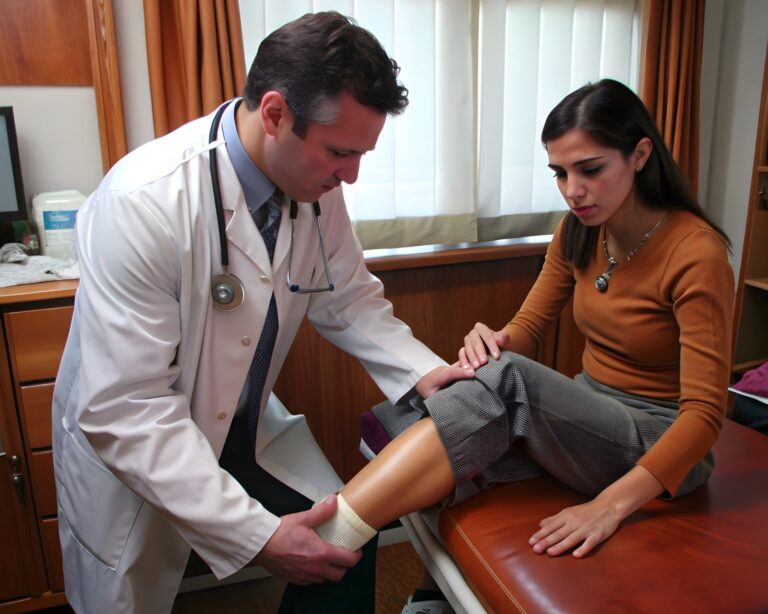

Yes, cartilage damage following an ACL tear can significantly impact surgical outcomes and recovery timelines. When the knee experiences a sudden twisting injury, both the ligament and cartilage surfaces can be damaged simultaneously. Cartilage injuries may slow healing, increase pain during rehabilitation, and elevate the risk of early-onset arthritis. However, modern surgical techniques, combined with targeted rehabilitation, can manage these complications effectively.
Quick Summary
- ACL tears often occur alongside cartilage injuries in the knee joint due to the same traumatic twisting mechanism
- Cartilage damage complicates recovery by extending rehabilitation timelines and increasing post-operative pain
- MRI and arthroscopy help identify cartilage injuries before surgery
- Surgical options like microfracture and chondroplasty can treat cartilage during ACL reconstruction
- Early surgery and structured physiotherapy protect remaining cartilage and improve long-term outcomes
- Weight management and muscle strengthening are critical post-surgery to reduce cartilage stress
- Return-to-sport testing ensures the knee is truly ready before resuming athletics
What Is Cartilage Damage After an ACL Tear?
Cartilage is a smooth, slippery tissue that covers the ends of bones inside the knee. It allows bones to glide smoothly during movement. When an ACL tear occurs from a sudden twisting injury, the forces that damage the ligament can also bruise, crack, or erode the cartilage surface. This is called articular cartilage damage or chondral injury. Unlike ligament tears, cartilage doesn’t heal well on its own because it has poor blood supply. Even small cartilage injuries can become larger over time if the knee remains unstable or is subjected to high-impact activities.
Why Does Cartilage Damage Happen During an ACL Injury?
When a person experiences a sudden twisting motion or direct impact to the knee, multiple structures are affected simultaneously. The ACL tear occurs when the ligament stretches beyond its limit or snaps completely. At the same time, the femur (thighbone) and tibia (shinbone) can shift abnormally, creating friction and impact forces on the cartilage surfaces. Beneath the cartilage, bone bruises (subchondral contusions) often occur, which can compromise cartilage healing.
Recent orthopedic research shows that cartilage damage is present in approximately 50–60% of acute ACL injuries. The severity depends on factors such as:
- Impact force: High-velocity injuries cause more damage
- Instability level: Greater ligament damage leads to more cartilage trauma
- Age: Younger athletes often have more cartilage damage because of higher-impact mechanisms
- Associated injuries: Meniscal tears (torn cartilage pads) often accompany ACL tears, compounding cartilage injury
Can Cartilage Damage Affect ACL Reconstruction Success?
Yes, cartilage damage can impact the success of ACL reconstruction in several ways:
Healing Complications: Cartilage does not regenerate naturally. Even after ACL surgery repairs the ligament, damaged cartilage cannot fully restore its smooth surface. This roughness increases friction during movement, potentially accelerating cartilage wear.
Pain Levels: Patients with cartilage damage often experience higher pain levels during and after rehabilitation. This can delay progression through physical therapy phases and slow the return to normal activities.
Long-Term Function: While ACL reconstruction is highly successful for restoring knee stability (success rates exceed 90%), the presence of cartilage damage may limit athletic performance and increase arthritis risk in later years. Studies indicate that cartilage damage present at the time of ACL surgery is one of the strongest predictors of early-onset knee osteoarthritis.
Success Metrics: Surgeons define success as restoring stability and pain-free function. Cartilage injuries can complicate both goals, requiring more aggressive rehabilitation protocols and careful activity management.
Does Cartilage Damage Delay Recovery After ACL Surgery?
Cartilage damage typically extends the recovery timeline. Here’s why:
Walking and Mobility: Patients with cartilage injury take longer to regain confident walking patterns. Swelling and inflammation from damaged cartilage surfaces persist longer than straightforward ligament injuries.
Rehabilitation Progression: Physical therapy must account for cartilage healing limitations. Therapists often recommend partial weight-bearing for longer periods and delayed introduction of high-impact activities like jumping or running.
Muscle Strength: Rebuilding quadriceps and hamstring strength is crucial, but cartilage sensitivity may limit early strengthening exercises. Recovery to full muscle strength can take 9–12 months instead of the typical 6–8 months.
Return to Sports: Athletes with cartilage damage may need 12–15 months to return to sport safely, compared to 9–12 months for isolated ACL tears. Sport-specific testing becomes even more critical to ensure cartilage tolerance.
What Are the Signs That Cartilage Damage May Be Present?
Several clinical indicators suggest cartilage injury alongside the ACL tear:
- Persistent swelling lasting weeks after the injury, even with ice and elevation
- Mechanical symptoms like knee locking (inability to fully straighten) or catching sensations during movement
- Grinding or popping sounds during knee bending, suggesting rough cartilage surfaces
- Localized pain in specific areas of the knee rather than diffuse pain
- Increased pain with weight-bearing activities beyond what is expected for a simple ACL tear
- Morning stiffness that improves gradually throughout the day
- Reduced range of motion compared to the uninjured knee, particularly in full extension or deep bending
Patients experiencing these signs should inform their orthopedic surgeon, as it affects surgical planning and post-operative expectations.
How Is Cartilage Damage Diagnosed?
Several diagnostic methods help identify cartilage injury:
MRI (Magnetic Resonance Imaging)
Advantages: MRI provides detailed images of soft tissues, including cartilage, bone bruises, and ligament tears. It’s non-invasive and highly sensitive for detecting cartilage defects.
Limitations: Not all cartilage injuries appear on MRI, especially very small surface-level damage. MRI cannot assess the true depth or severity of damage until the joint is directly visualized.
Clinical Examination
Advantages: Physical examination by an experienced orthopedic surgeon can identify mechanical symptoms (locking, catching) and pain patterns that suggest cartilage injury.
Limitations: Clinical tests are subjective and may miss cartilage injuries that don’t produce obvious mechanical symptoms.
Arthroscopy
Advantages: Direct visualization of the knee joint interior via a small camera. Arthroscopy is the gold standard for detecting and treating cartilage damage. Surgeons can assess damage depth, location, and size precisely.
Limitations: Arthroscopy is invasive and requires anesthesia. It’s typically reserved for surgical intervention rather than diagnosis alone, though diagnostic arthroscopy is sometimes performed before full reconstruction surgery.
Can Cartilage Damage Be Treated During ACL Reconstruction?
Yes, cartilage injuries can be addressed during the same ACL reconstruction procedure:
Microfracture Technique
This technique deliberately creates small holes in the bone beneath damaged cartilage, stimulating the body’s natural healing response. Bleeding from bone marrow cells fills the defect and develops fibrocartilage (scar-like cartilage). This is effective for small defects (less than 2 cm²) and can improve pain and function.
Chondroplasty
Surgeons smooth rough, irregular cartilage surfaces to reduce friction. While this doesn’t repair damage, it can decrease pain and mechanical symptoms caused by rough cartilage surfaces.
Cartilage Restoration Procedures
For larger cartilage defects, surgeons may use advanced techniques such as:
- Autologous chondrocyte implantation (ACI): Growing cartilage cells in a laboratory and implanting them into the defect
- Osteochondral autograft transfer: Moving healthy cartilage and bone from a non-weight-bearing area to the damaged zone
These techniques are more complex and typically performed in specialized centers.
Combined Procedures
Most commonly, surgeons address both the ACL tear and cartilage damage in a single surgery, allowing coordinated healing and reducing the need for multiple operations.
Does Early Surgery Protect Knee Cartilage?
Current evidence suggests that early ACL reconstruction may offer cartilage protection benefits:
Stability Restoration: Delaying ACL surgery leaves the knee mechanically unstable. This instability increases abnormal knee motion, creating repeated micro-trauma to cartilage surfaces. The longer the instability persists, the greater the cumulative cartilage damage.
Repeated Injury Risk: An unstable knee is vulnerable to re-injury from simple activities like stepping on uneven ground or making cutting motions during sports. Each subsequent injury can cause additional cartilage damage, compounding the initial injury.
Inflammation Control: Early surgery, combined with prompt rehabilitation, helps control chronic inflammation in the joint. Prolonged inflammation accelerates cartilage degeneration.
Research Findings: Studies comparing early surgery (within 3 months of injury) to delayed surgery show that early intervention is associated with lower rates of secondary meniscal tears and cartilage progression. However, the relationship between surgery timing and long-term cartilage outcomes is complex and depends on many individual factors.
What Can Patients Do to Improve Recovery?
Evidence-based strategies enhance outcomes after ACL reconstruction, especially when cartilage damage is present:
Physiotherapy and Rehabilitation
Structured physical therapy is fundamental. Therapists use protocols specifically designed for ACL reconstruction that progress strength and proprioception systematically. When cartilage damage is present, therapists adjust loading progressions to protect healing cartilage.
Muscle Strengthening
Robust quadriceps and hamstring strength protects the knee by improving stability and reducing abnormal joint motion. Strong muscles absorb forces that might otherwise stress cartilage. Studies show that quadriceps strength deficits significantly increase cartilage wear over time.
Weight Management
Excess body weight increases compressive forces on knee cartilage. For every 1 kg of body weight gained, the knee experiences approximately 4 kg of additional compressive force during walking. Even modest weight loss can substantially reduce cartilage stress, especially important in the months following surgery.
Activity Modification
Avoiding high-impact activities (running, jumping, cutting sports) during early recovery protects healing cartilage. Gradual, progressive return to higher-impact activities, guided by a physical therapist, reduces re-injury risk and cartilage re-damage.
Return-to-Sport Testing
Before returning to competitive activities, athletes should complete objective testing including:
- Strength testing (ensuring 90% strength symmetry between knees)
- Hop testing (single-leg and triple-hop distances)
- Agility drills and cutting tests
- Sport-specific skill assessments
These tests ensure the knee is biomechanically ready and reduces cartilage re-injury risk.
Comparison Table: ACL Tear With Cartilage Damage vs ACL Tear Without Cartilage Damage
| Feature | ACL Tear Only | ACL Tear + Cartilage Damage |
| Initial Pain Level | Moderate to severe | Severe, often with mechanical symptoms |
| Early Swelling | Subsides within 2-4 weeks | Persists 4-8 weeks or longer |
| Recovery Timeline | 9-12 months | 12-15 months or more |
| Surgery Type | Standard ACL reconstruction | ACL reconstruction + cartilage treatment |
| Arthritis Risk (10 years) | 15-25% | 40-60% |
| Physical Therapy Duration | 6-8 months | 9-12 months |
| Return to Full Sport | 9-12 months | 12-15 months |
| Long-Term Function | Excellent in most patients | Good, but cartilage-dependent |
Signs You Should See an ACL Specialist
Several indicators warrant consultation with an orthopedic surgeon specializing in sports injuries:
- Knee gives way or feels unstable during walking or activities
- Persistent swelling that doesn’t improve with rest and ice
- Mechanical symptoms (locking, catching, popping) that interfere with daily life
- Pain that limits stairs, squatting, or returning to work
- Injury mechanism involving sudden pivoting or twisting motions
- Significant pain or swelling after the initial acute phase has passed
- Uncertainty about whether surgery is necessary for your specific injury
When Should You Consult an Orthopedic Surgeon?
Follow this timeline for proper evaluation:
- Immediately – Seek urgent care if the knee gives way or severe pain/swelling develops; rule out emergencies
- Within 1 week – Schedule consultation with orthopedic surgeon for formal evaluation and imaging if not already done
- Before 3 weeks – Complete diagnostic imaging (X-rays, MRI) to assess ligament and cartilage damage
- By 4-6 weeks – Decide on surgical vs. non-surgical treatment based on imaging findings and functional goals
- If surgery planned – Schedule ACL reconstruction ideally within 3 months to limit secondary damage
- Post-surgery – Begin structured rehabilitation immediately and commit to 9-12+ months of focused therapy
- At 6-9 months – Assess readiness for sport-specific training with your physical therapist
- At 12 months – Perform return-to-sport testing before resuming high-impact athletics
FAQ: Patient Questions About Cartilage Damage and ACL Recovery
Can cartilage heal after an ACL tear?
Cartilage has very limited healing capacity due to its poor blood supply. Minor cartilage injuries may stabilize, but they don’t repair completely. Surgical techniques like microfracture can stimulate fibrocartilage formation, which provides functional improvement but doesn’t restore original cartilage. Managing the injury to prevent progression is the realistic goal.
Does cartilage damage mean I automatically need surgery?
Not necessarily for the cartilage injury itself. However, most ACL tears require surgery to restore knee stability. If cartilage damage is present, it becomes one factor in discussing surgical timing and technique. Your surgeon will recommend the approach that best protects remaining cartilage.
Can MRI detect all cartilage damage?
MRI detects cartilage defects well but may miss very small surface irregularities. Arthroscopic visualization during surgery is the definitive method for assessing cartilage damage. However, MRI typically provides sufficient information for surgical planning in most cases.
Will cartilage damage make my recovery take much longer?
Yes, cartilage damage usually extends recovery by 2-4 months compared to isolated ACL tears. The timeline depends on damage extent, your age, and how well you follow rehabilitation protocols. Commitment to physiotherapy is essential for optimizing outcomes.
Can cartilage damage cause arthritis after ACL surgery?
Cartilage damage present at the time of ACL surgery increases arthritis risk. Studies show patients with cartilage injuries have higher osteoarthritis rates at 10+ years. However, this doesn’t mean arthritis is inevitable—proper rehabilitation, weight management, and activity modification help minimize risk.
Is cartilage repaired during ACL surgery?
Techniques like microfracture and chondroplasty address cartilage during ACL reconstruction. However, “repair” is misleading—these techniques stimulate fibrocartilage formation or smooth surfaces rather than restoring original cartilage. The goal is functional improvement, not complete restoration.
Can athletes return to competitive sports after ACL and cartilage injury?
Yes, many athletes successfully return to sport, but expectations may differ from those with isolated ACL tears. Return-to-sport testing is especially important. Some athletes modify sport intensity or position to reduce joint stress. Open discussion with your surgeon and physiotherapist about realistic goals is essential.
How can I protect my knee after surgery if cartilage is damaged?
Weight management is critical—excess weight accelerates cartilage wear. Strong leg muscles provide dynamic stability and protection. Avoid high-impact activities that stress healing cartilage. Graduated return to activity, following your physiotherapist’s guidance, protects the repair. Regular check-ups help monitor cartilage progression.
Expert Insight: Dr. Abhinandan Punit on ACL and Cartilage Management
Dr. Abhinandan Punit is a European Board Certified Orthopedic Surgeon and Sports Medicine Specialist with 15+ years of experience managing complex knee injuries in Bangalore. With 1,000+ orthopedic procedures performed annually and over 300 sports medicine cases, Dr. Punit brings deep expertise to ACL reconstruction and cartilage injury management.
At Elite Orthocare & Multi Speciality Clinic on Kanakapura Road and Narayana Health City in Electronic City, Dr. Punit employs advanced diagnostic techniques and minimally invasive surgical approaches. His clinical approach emphasizes early intervention to prevent secondary cartilage damage and customized rehabilitation protocols that account for cartilage injuries.
“When a patient presents with an ACL tear and cartilage damage, our goal is twofold: restore ligament stability while protecting remaining cartilage health,” explains Dr. Punit. “Early, accurate diagnosis through MRI and selective arthroscopy, combined with surgical expertise and committed physiotherapy, gives patients the best chance of returning to their desired activities with durable long-term outcomes.”